

Immunohistochemistry (IHC) is a specialised technique that works on the principle of antigen-antibody reaction. The antigen in the tissue sample is detected by using specific antibodies and the binding of these is detected under a microscope / whole slide image. It is used to diagnose a disease, help in choice of therapy, monitor response to therapy and in predicting prognosis.
Triesta Sciences has highly experienced subspecialist pathologists who use relevant antibodies to accurately diagnose or monitor the disease. Strict quality control practices including participation in proficiency testing programs ensure accurate reports.
Services Offered:
- Frozen section
- Subspecialty Histopathology for Oncology and Non-Oncology cases
- Immunohistochemistry (IHC)
- Digital Pathology with machine learning algorithm
- Cytopathology including LBC and Cell block.
- Secondary consults for reporting on digital images (WSI)
- Collaboration for clinical research and biomarker research projects, AIML projects
- We also offer an IHC slide staining facility.
USP:
- 200+ IHC Markers including companion diagnostic markers
- Experienced and highly competent histopathologists with subspecialty expertise and reporting as below
➣ CNS
➣ Head & Neck
➣ Breast
➣ Lung & Thoracic organ
➣ Gastrointestinal
➣ Hepatobiliary
➣ Female Reproductive System
➣ Urogenital
➣ Bone & Soft Tissue
➣ Derma
➣ Pediatric
➣ Endocrine Organs
➣ Lymphoreticular system
➣ Eye and Ocular Adnexal
- Secondary consults on whole slide images
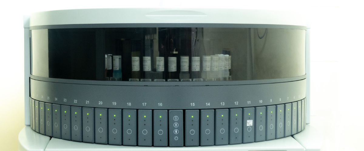
- Fully automated FDA approved IHC systems.
- Companion diagnostic markers for all three clones of PDL1 (22c3, SP263 & SP142)
- Comprehensive panels for complete workup – Sarcoma, CUP, Lymphoma Panel etc
- Organ & disease specific panels – Breast, Lung, Colorectal, Hematolymphoid, Endometrioid, H&N etc
- MMR / MSI panel
- In-Situ-Hybridization (ISH) for EBV, Kappa and Lambda Light chains
- Immunotherapy & TILs panel
- DISH for Her-2/neu Equivocal cases
- Research collaboration for drug discovery and newer biomarkers development.
Most Frequently asked Tests
FAQ
How should I bring the biopsy samples to the lab for testing?
The biopsy sample has to be preserved in a solution ‘10% Buffered Neutral Formalin’, in a quantity enough to completely immerse the sample. The sample can be delivered to the laboratory with a proper label and the test request form from your doctor. Try to submit the sample within 2-3 days to the laboratory.
Can my biopsy sample be preserved for further testing?
Representative tissue from the biopsy sample will be processed and preserved in wax / paraffin blocks. They can be preserved at room temperature for 5-10 years.
Can I meet the Pathologist to discuss my report?
Our Pathologists are available at the laboratory during the working hours for any discussion on the reports. The customer care team can be contacted with a request to speak to the pathologists.
What is the frozen section test?
This is a test performed on fresh operated specimens, when the patient is still under anaesthesia, on the OT table. This test helps the surgeon to decide on the margin status of his surgery and plan extending the surgery to achieve complete removal of the tumour.
When will Immunohistochemistry test be recommended
Immunohistochemistry (IHC) is a specialised test done on tissue samples in paraffin blocks. They are recommended to,
- Diagnose a disease when there is ambiguity in histopathology.
- Determine prognosis.
- Decide on choice of treatment.
- Predict treatment response.
- Monitor treatment response.
Can the IHC test be performed on the wax blocks?
Yes. It can be performed provided the blocks are well preserved.
Can I get the digital images of my biopsy sample?
Yes. Digital images will be sent over an email upon request to our customer care team.
How is FNAC different from Biopsy?
FNAC is an outpatient procedure, where cells are aspirated from the suspicious areas in the body either directly or under ultrasound / CT scan guidance. This is done by Pathologists or the radiologists. While biopsy is usually done under anaesthesia, and a good chunk of tissue is removed by the radiologist, or the entire lesion is removed by the surgeon. FNAC reports are usually available in a day or two, the biopsy report may take 2-10 days, depending on the type of sample.
When will I get the Histopathology report?
Please ask our customer care executive, as the turnaround time varies depending on the sample type.
Can the Histopathology test be performed without a doctor’s recommendation?
It is recommended to get a proper request form from your doctor along with the specimen for histopathology test. However, reviews or opinions can be provided on paraffin blocks / slides at your request.