

The Department of Histopathology at Triesta Science includes Routine Histopathology, Cytopathology, Immunohistochemistry and Digital Pathology. One of the largest departments within the lab handling more than 30,000 patient samples a year from our internal and external customers with subspecialty reporting by a team of pathologists.
Diagnosis is made by looking at tissue samples under a microscope or digital whole slide image. Some of the cases require additional tests like Immunohistochemistry or ISH to aid in diagnosis and provide information on therapeutic or prognosis. We offer customized case work-up for diagnosis and treatment choices, by collaborating with clinical doctors / surgeons / oncologists on a day-to-day basis. In addition, Frozen section services are available to both in-house and external patients on OT table for rapid diagnosis.
The department is well equipped with advanced technology including the FDA approved platform for Immunohistochemistry and Digital Pathology.
The department has highly competent professional and technical staff, residents, researchers with equal focus on both cancer and non-cancer cases affecting adults and pediatric patients.
The team is actively involved in research and developing newer diagnostic tests to improve diagnosis, monitoring, and treatment options. Stringent quality control protocols to ensure optimum tissue handling. Participation in International Proficiency Testing programs with excellent results each time.
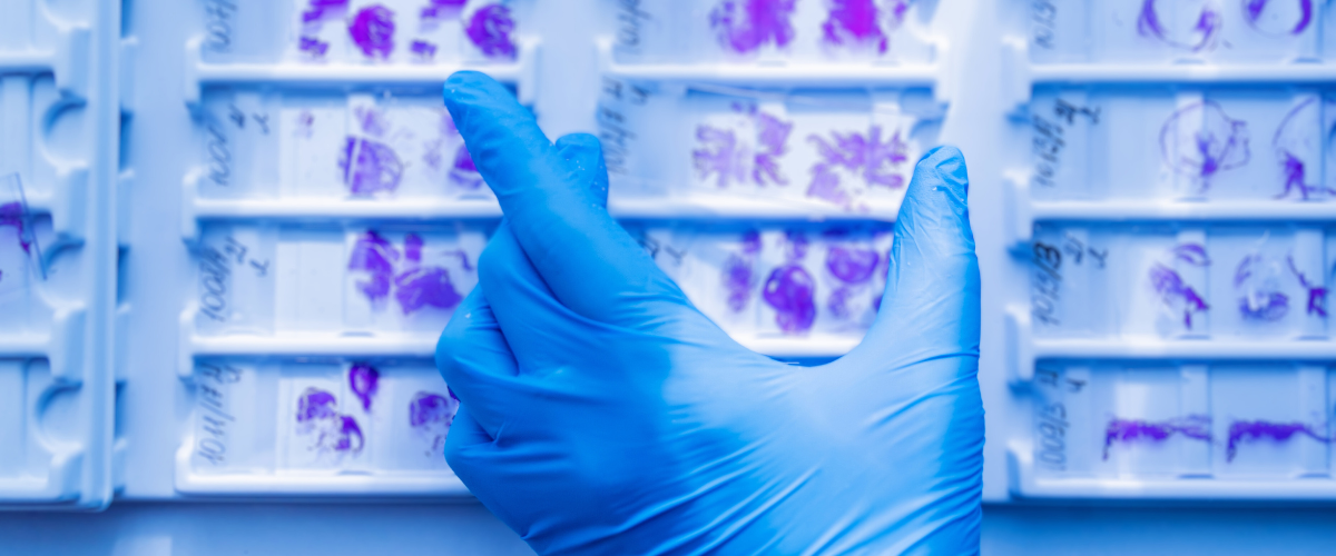
Digital Pathology
The latest technology in converting a glass slide with a tissue section to a whole slide high quality high resolution digital image that can be accessed and viewed across the world.
Triesta Sciences is the first lab in India to install digital scanners. The technology has enabled us to provide / establish network digital pathology practice across our centers in India and abroad.
Recipient of ‘first 100% digital pathology lab for primary diagnosis’ award in 2019.
Specialization:
- IVD-CE approved breast biomarker algorithm for quantitative interpretation
- National / International consultation for expert opinion
- Digital image archive and library for education and ML projects
- Members of Digital Pathology Association, USA
- Numerous publications on digital pathology in international journals
- Participate in development and validation of many Machine Learning Algorithms
- Offer annotation and slide scanning services.
CYTOPATHOLOGY
The cells are obtained by minimally invasive procedures that can be analyzed for diagnosis or treatment monitoring e.g. FNAC / scrapings / brushes as in Pap smear else from sediments of body fluids including Urine and CSF. The subspecialist pathologists in the department perform procedures as above of their area of specialization (organ specific) and report the cases with accuracy.
Specialization:
- Fine Needle Aspiration Cytology (FNAC)
- Pap Smears and LBCs
- Cell block for all body fluids
- Reporting on EBUS, TBNA and Biliary Brush Cytology by subspecialist pathologist.
- ROSE for hospital cases
Services Offered:
- Frozen section
- Immunohistochemistry (IHC)
- Digital Pathology with machine learning algorithm
- Cytopathology including LBC and Cell block.
- Secondary consults for reporting on digital images (WSI)
- Collaboration for clinical research and biomarker research projects, AIML projects
- IHC slide staining facility.
USP:
- Experienced and highly competent histopathologists
- Subspecialty reporting as below by a dedicated clinical team.
➣ CNS
➣ Head & Neck
➣ Breast
➣ Lung & Thoracic organ
➣ Gastrointestinal
➣ Hepatobiliary
➣ Female Reproductive system
➣ Urogenital
➣ Bone & Soft Tissue
➣ Derma
➣ Pediatric
➣ Endocrine Organs
➣ Lymphoreticular system
➣ Eye and Ocular Adnexal
- Secondary consults on whole slide images
Equipment:
- FDA approved digital pathology scanners.
- Fully automated FDA approved IHC equipment.
- IVD-CE approved machine learning algorithm for breast biomarker interpretation
- Automated film coverslip machine
- Automated stainer, microtomes, tissue embedding stations.
- Automated tissue processors
- Cryostat
- Cytospin and Eziprep
- Automated bone decalcifier
Most Frequently asked Tests
FAQ
How should I bring the biopsy samples to the lab for testing?
The biopsy sample has to be preserved in a solution ‘10% Buffered Neutral Formalin’, in a quantity enough to completely immerse the sample. The sample can be delivered to the laboratory with a proper label and the test request form from your doctor. Try to submit the sample within 2-3 days to the laboratory.
Can my biopsy sample be preserved for further testing?
Representative tissue from the biopsy sample will be processed and preserved in wax / paraffin blocks. They can be preserved at room temperature for 5-10 years.
Can I meet the Pathologist to discuss my report?
Our Pathologists are available at the laboratory during the working hours for any discussion on the reports. The customer care team can be contacted with a request to speak to the pathologists.
What is the frozen section test?
This is a test performed on fresh operated specimens, when the patient is still under anaesthesia, on the OT table. This test helps the surgeon to decide on the margin status of his surgery and plan extending the surgery to achieve complete removal of the tumour.
When will Immunohistochemistry test be recommended
Immunohistochemistry (IHC) is a specialised test done on tissue samples in paraffin blocks. They are recommended to,
- Diagnose a disease when there is ambiguity in histopathology.
- Determine prognosis.
- Decide on choice of treatment.
- Predict treatment response.
- Monitor treatment response.
Can the IHC test be performed on the wax blocks?
Yes. It can be performed provided the blocks are well preserved.
Can I get the digital images of my biopsy sample?
Yes. Digital images will be sent over an email upon request to our customer care team.
How is FNAC different from Biopsy?
FNAC is an outpatient procedure, where cells are aspirated from the suspicious areas in the body either directly or under ultrasound / CT scan guidance. This is done by Pathologists or the radiologists. While biopsy is usually done under anaesthesia, and a good chunk of tissue is removed by the radiologist, or the entire lesion is removed by the surgeon. FNAC reports are usually available in a day or two, the biopsy report may take 2-10 days, depending on the type of sample.
When will I get the Histopathology report?
Please ask our customer care executive, as the turnaround time varies depending on the sample type.
Can the Histopathology test be performed without a doctor’s recommendation?
It is recommended to get a proper request form from your doctor along with the specimen for histopathology test. However, reviews or opinions can be provided on paraffin blocks / slides at your request.